Untitled

Human Reproduction Vol.23, No.2 pp. 324–328, 2008 Advance Access publication on December 11, 2007 Obesity affects spontaneous pregnancy chancesin subfertile, ovulatory women Jan Willem van der Steeg1,2,4,8, Pieternel Steures1,2,4, Marinus J.C. Eijkemans1,J. Dik F. Habbema1, Peter G.A. Hompes4, Jan M. Burggraaff5, G. Jur E. Oosterhuis6,Patrick M.M. Bossuyt3, Fulco van der Veen2 and Ben W.J. Mol2,7 1Department of Public Health, Erasmus MC, University Medical Centre Rotterdam, The Netherlands; 2Center for Reproductive Medicine,Academic Medical Centre, Amsterdam, The Netherlands; 3Department of Clinical Epidemiology and Biostatistics, Academic MedicalCentre, Amsterdam, The Netherlands; 4Department of Obstetrics and Gynaecology, Vrije Universiteit Medical Centre, Amsterdam,The Netherlands; 5Department of Obstetrics and Gynaecology, Scheperziekenhuis Emmen, Emmen, The Netherlands; 6Departmentof Obstetrics and Gynaecology, Medisch Spectrum Twente, Enschede, The Netherlands; 7Department of Obstetrics and Gynaecology,Ma´xima Medical Centre, Veldhoven, The Netherlands 8Correspondence address. Academic Medical Centre, Centre for Reproductive Medicine, Room H4-213, PO Box 22660, 1105 AZAmsterdam, The Netherlands. Tel: þ31-20-5663857; Fax: þ31-20-6963489; E-mail: [email protected] BACKGROUND: Obesity is increasing rapidly among women all over the world. Obesity is a known risk factor forsubfertility due to anovulation, but it is unknown whether obesity also affects spontaneous pregnancy chances in sub-fertile, ovulatory women. METHODS: We evaluated whether obesity affected the chance of a spontaneous pregnancyin a prospectively assembled cohort of 3029 consecutive subfertile couples. Women had to be ovulatory and had tohave at least one patent tube, whereas men had to have a normal semen analysis. Time to spontaneous ongoing preg-nancy within 12 months was the primary endpoint. RESULTS: The probability of a spontaneous pregnancy declinedlinearly with a body mass index (BMI) over 29 kg/m2. Corrected for possible related factors, women with a high BMIhad a 4% lower pregnancy rate per kg/m2 increase [hazard ratio: 0.96 (95% CI 0.91 – 0.99)]. CONCLUSIONS: Theseresults indicate that obesity is associated with lower pregnancy rates in subfertile ovulatory women.
Keywords: obesity; subfertility; pregnancy chance; spontaneous conception 25% in North America (Butler, 2004; Linne´, 2004; Haslam and The use of assisted fertility techniques has increased James, 2005; Watson, 2005). The main adverse consequences tremendously in the past three decades (Society for Assisted are cardiovascular disease, type 2 diabetes and cancer. Overall, Reproductive Technology and American Society for Repro- it is thought to be the sixth most important risk factor for mor- ductive Medicine, 2002). Although this may be due to better tality and morbidity (Allison et al., 1999). Furthermore, obesity availability of the wide range of current technologies, an is a known risk factor for anovulation, which may lead to subfer- increased demand for fertility care may play a role as well.
tility (Rogers and Mitchell, 1952; Hartz et al., 1979; Norman and This increased demand may be due to an increased incidence of Chlamydia trachomatis and increased maternal age Current NICE fertility guidelines recommend that all obese (Martin, 2000; Coombes, 2004). Additionally, obesity is women, regardless of their cycle characteristics, should be expected to be a potential cause for an increase in subfertility informed that they are likely to take longer to conceive in the near future (Bolu´mar et al., 2000).
(National Institute for Clinical Excellence, 2004). This rec- Obesity is increasing rapidly all over the world, affecting more ommendation is based on three studies. Two studies analysed than one billion people worldwide (Haslam and James, 2005).
the relationship between BMI and time to pregnancy in The World Health Organization (WHO) considers a body mass women who were pregnant or had delivered a child (Jensen index (BMI) as abnormal if BMI is over 25.0 kg/m2 and et al., 1999; Bolu´mar et al., 2000), whereas the third study ana- defines obesity as a BMI over 30.0 kg/m2 (World Health Organ- lysed fat distribution and the chance of conceiving in women in isation, 1995). More women of reproductive age are becoming a donor insemination programme (Zaadstra et al., 1993). All overweight and obese. Nowadays, the incidence of obesity in studies reported a negative effect of obesity on the chance of women of child bearing age is 12% in Western Europe and pregnancy in these potentially fertile women.
The Author 2007. Published by Oxford University Press on behalf of the European Society of Human Reproduction and Embryology.
All rights reserved. For Permissions, please email: [email protected] However, evidence that obesity also affects the chance of spontaneous pregnancy in subfertile ovulatory women is still We first assessed the relation between BMI and probability of preg- lacking. The aim of this study was to determine whether nancy through spline functions. By visual inspection, it was deter- obesity in subfertile ovulatory women is associated with a mined whether BMI behaved as a linear or non-linear function in decreased chance of spontaneous pregnancy.
relation to the probability of spontaneous pregnancy, and whethercut-off values for optimal BMIs could be observed. Non-linearitywas tested with ANOVA analysis (Harrell et al., 1988).
We then analysed the predictive capacity of BMI as hazard ratios (HR) by Cox proportional hazard analysis of the time to spontaneous Between January 2002 and February 2004, we included consecutive ongoing pregnancy. The proportional hazards assumption was evalu- subfertile couples that had not been evaluated previously for subferti- ation with S-plus (Grambsch and Therneau, 1994). Lastly, we repeated lity, in a prospective cohort study. The study was performed in 24 hos- the analysis correcting for possibly related factors in a multivariable pitals in the Netherlands. The detailed study protocol has been hazards regression model. Potential related factors were female age, documented in a previous publication (van der Steeg et al., 2007).
duration of subfertility, previous pregnancy, referral status, semen In short, all couples underwent a fertility work-up consisting of: a fer- motility and current smoking of the female and male partner tility history, including details about height and weight, smoking habits, (Bolu´mar et al., 2000; Hunault et al., 2004).
assessment of ovulation, assessment of tubal patency and semen analy- In all analyses, a P-value of 0.05 was used to indicate significance.
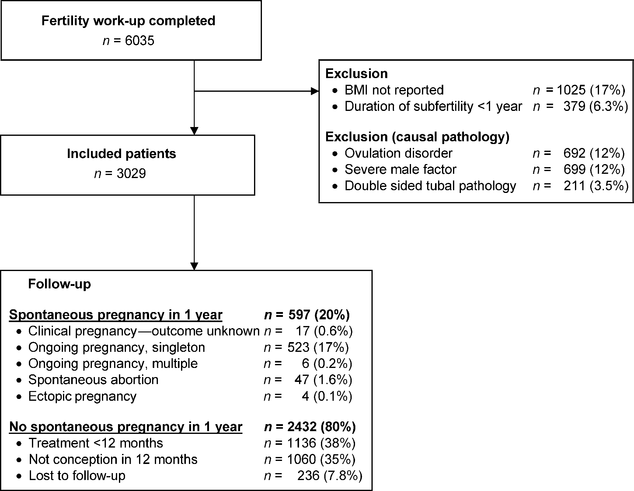
sis (Dutch Society of Obstetrics and Gynaecology, 2004). Duration of Calculations were performed with SPSSw 12.0 (SPSS Inc., Chicago, subfertility and female and male age were set at the end of the infertility IL, USA) and S-plusw 6.0 (MathSoft Inc., Seattle, WA, USA) assessment. Subfertility was considered to be secondary if a woman had conceived in the current or in a prior partnership, regardless of the preg-nancy outcome. The BMI was calculated as the weight in kilogramsdivided by the square of the height in meters, both self-reported during the first visit. BMI of the men as well as timing and frequency The study was approved by the local ethics committee of each partici- Fertility work-up of the female partnerOvulation was assessed by means of a basal body temperature chart, measurement of mid-luteal serum progesterone or by sonographic In total, 6035 subfertile couples that had completed their ferti- monitoring of the cycle. The menstrual cycle was considered regular lity work-up were registered. Of these, 379 (6.3%) couples if the duration of the cycle was between 23 and 35 days, with an inter- with a duration of subfertility less than 1 year, 692 (12%) cycle variation of less than 8 days, during the past year (Munster et al., couples with anovulation, 211 (3.5%) couples with two-sided 1992). Tubal pathology was assessed by a chlamydia antibody test tubal pathology and 699 (12%) couples with severe male (CAT), hysterosalpingography (HSG) or laparoscopy. Those with a factor were excluded (Fig. 1). In 1025 (17%) couples the BMI positive CAT subsequently went on to have further investigationwith a HSG or laparoscopy (Mol et al., 1997). Couples, in whom was not reported. Therefore, 3029 couples were included in the female partner was diagnosed with anovulation or with two-sided the analysis. Follow-up was completed for 2793 couples tubal pathology, were excluded from the analysis.
(92%). Of all couples, 529 (17%) had a spontaneous ongoingpregnancy within 1 year (Fig. 1). In 17 women, pregnancy outcome was unknown. There were 47 women (7.8% of all Semen analysis was performed at least once according to the WHO pregnancies) who miscarried and four women (0.7% of all preg- guidelines, including semen volume, concentration, morphology and nancies) who had an ectopic pregnancy. Within 12 months, motility (World Health Organisation, 1999). Couples in whom theman had a total motile sperm count (TMC) ,3 Â 106 were excludedfrom the study.
Follow-upAfter completion of the fertility work-up, the probability of a spon-taneous pregnancy within 1 year, leading to live birth, was calculatedwith a validated prediction model (http://www.freya.nl/probability.
php) (Hunault et al., 2004; van der Steeg et al., 2007). Dependingon that probability couples were counselled for expectant managementor fertility treatment according to the national fertility guidelines(Dutch Society of Obstetrics and Gynaecology, 1998; Dutch Societyof Obstetrics and Gynaecology, 2000). The exact study flow hasbeen reported in a previous paper (van der Steeg et al., 2007).
Couples were followed prospectively from the completion of the fer-tility work-up until pregnancy or start of treatment within 12 months.
The primary endpoint was time to conception without treatment,resulting in an ongoing pregnancy and counted in calendar timeused in a continuous way. Couples who did not conceive were cen-sored when treatment started or at the last date of contact duringfollow-up.
1136 (38%) couples started treatment, whereas 1060 (35%) 29 kg/m2 (95% CI 0.91 – 0.99)]. A BMI below 21 kg/m2 was neither started treatment nor became pregnant. Median length associated with a lower probability of spontaneous ongoing of follow-up was 28.1 weeks (fifth to 95th percentile: 2 – 134 pregnancy than the reference group, but was not statistically weeks), 18.5 weeks (1 – 100 weeks) for those who conceived significant [HR 0.97 per kg/m2 below 21 kg/m2 (95% CI and 31.5 weeks (2 – 146 weeks) for those without a pregnancy.
Baseline characteristics are represented in the Table I. The median BMI was 22.9 kg/m2 (5–95th percentile: 19–33 kg/m2).
A BMI below 18.5 kg/m2 was found in 3.7% of the women,between 18.5 and 25 in 67%, between 25 and 30 in 19%,between 30 and 35 kg/m2 in 6.7%, and !35 kg/m2 in 3.8%.
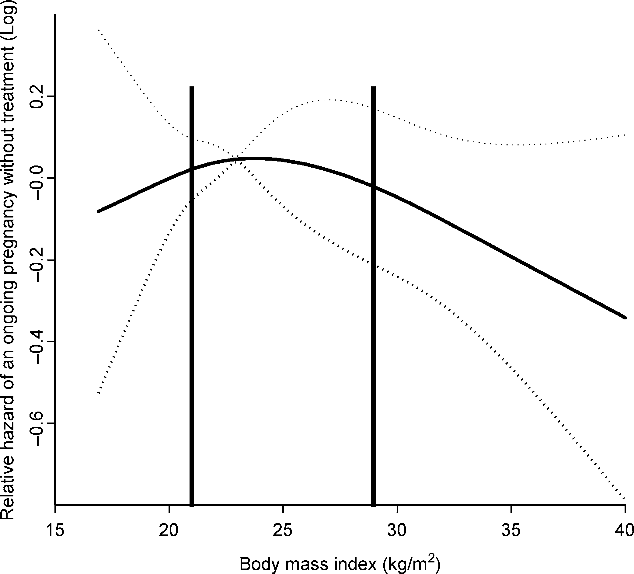
Couples, in whom the BMI was not documented, were onaverage older, more often secondary subfertile and moreoften referred by a gynaecologist (ANOVA statistics,P , 0.05), although differences were small. Other baselinecharacteristics were comparable between the groups. Thespline analysis showed that BMI had an inversed U-shapedrelationship with the probability of pregnancy, although thiswas not statistically significant over the whole range (Fig. 2)(ANOVA P ¼ 0.4). From this spline function, two thresholdswere derived at 21 and 29 kg/m2. Women with a BMIbetween 21 and 29 were defined as the reference group. Theunivariable analysis showed that BMI, female age, durationof subfertility, secondary subfertility, referral status andsemen motility were statistically significantly related to theprobability of a spontaneous ongoing pregnancy (Table II).
A BMI above 29 kg/m2 was associated with a statistically Figure 2: Spline function of the BMI in relation to time to spon- significant lower probability of spontaneous ongoing preg- taneous ongoing pregnancy.
Relative hazardHR. Dotted lines represent 95% confidence inter- nancy than the reference group [HR 0.95 per kg/m2 above vals. Vertical lines show the thresholds of 21 and 29 kg/m2. BMIsabove 29 kg/m2 were significantly associated with a decreasedfecundity, whereas there was a trend below 21 kg/m2 Table II. Results of the univariable and multivariable Cox’ regressionanalysis.
aDifference in baseline characteristics between women with and without data on BMI, tested with ANOVA.
bTMC, Total motile sperm count.
aWomen with a BMI 21–29 kg/m2 were used as reference group.
0.87 – 1.07)] (Table II). The proportional hazards assumption, were small. However, we may have selected a group here that necessary to perform the Cox analysis in a correct way, was was more fertile than the overall group.
We can only speculate about the pathophysiological expla- The multivariable analysis, adjusted for female age, duration nations for the lower pregnancy chances in obese women. It of subfertility, previous pregnancy, referral status (second or has been suggested that leptin may be of importance (Rosen- third care), semen motility and current smoking of the female baum and Leibel, 1999; Mantzoros, 2000; Chan and Mantzoros, and male partner, did not change the results [HR 0.96 per 2005). Genetically mediated states of leptin deficiency result in kg/m2 (95% CI 0.91 – 0.99)]. In case of a woman with a BMI obesity and subfertility (Rosenbaum and Leibel, 1999).
of 35 kg/m2, the probability of spontaneous pregnancy was Decreasing leptin levels due to starvation result in decreased 26% lower, and in case of a woman with a BMI of 40 kg/m2, estradiol levels and amenorrhoea (Mantzoros, 2000). There is it was 43% lower compared with women with a BMI evidence that leptin may influence ovarian steroidogenesis directly. Further research of the role of intra-ovarian leptinaction in relation to subfertility remains of interest.
It could be hypothesized that lifestyle interventions that focus on weight reduction are an effective intervention (Knowler This cohort study showed that obesity is an important risk et al., 2002). This study focused on the BMI at start of the fer- factor for pregnancy chances in subfertile, ovulatory women.
tility work-up, rather than on weight changes. In subfertile ano- For every BMI unit above 29 kg/m2, the probability was vulatory women, several studies have reported such a beneficial reduced by 5%, being a reduction comparable to the incre- effect (Hollmann et al., 1996; Pasquali et al., 1997; Crosignani ment of one year in female age. Given the increased prevalence et al., 2003). A next step could be to randomly allocate obese of obesity, this is a worrying finding.
and subfertile, ovulatory women to a controlled low-calories Up till now, the relationship between BMI and pregnancy diet, or to their normal diet and compare HR.
chances had not been established in ovulatory subfertile women.
In conclusion, the results of this study indicate that ovulatory This is the first prospective cohort study to demonstrate this. It subfertile women with a BMI over 29 kg/m2 have lower preg- differs from previous studies on obesity and pregnancy chances nancy rates compared with those with normal weight. Now, we in two ways. First, all other studies dealt with proven fertile popu- know that not only obese women with anovulation have lower lations, whereas our study included subfertile women. Second, chances of conception, but also obese women with a regular many studies dealt with obesity as a categorical variable. In con- cycle. Owing to the fact that more women of child-bearing trast, in this study, BMI was analysed as a continuous variable age are becoming overweight and obese, this is a worrying that allowed a subtle decline in pregnancy rate starting at 29 kg/m2 to be demonstrated. In proven fertile women, BMIwas reported to be a risk factor for the chance of conception inthe category of women with a BMI over 25 kg/m2 [HR 0.77 (95% CI 0.70 – 0.84) Jensen et al., 1999]. In the category of Ben W.J. Mol, Fulco van der Veen and Peter G.A. Hompes women with a BMI over 30 kg/m2, BMI was reported to be a designed the study. Jan Willem van der Steeg and Pieternel risk factor of having a delayed conception with an odds ratio Steures promoted it, co-ordinated this cohort study, collected of 12 (95% CI 3.7 – 36) (Bolu´mar et al., 2000).
the data and sought ethical approval. Jan M. Burggraaff and A limitation of this study is that frequency of intercourse was not Jur G.J.E. Oosterhuis included couples and collected data.
taken into account. Recently, a review found support that obesity Jan Willem van der Steeg did the analysis, under the super- is associated with decreased intercourse frequency, reduced vision of Ben W.J. Mol and Marinus J.C. Eijkemans. J. Dik sexual desire and erectile dysfunction (Larsen et al., 2007).
F. Habbema and Patrick M.M. Bossuyt provided statistical However, in view of the paucity of data, confounding factors advice. All authors helped to prepare the final report. Other like medication and adverse lifestyles could not be ruled out.
contributors in this multicentre study included couples and Another limitation of our study is the fact that the BMI of the are mentioned as a part of the CECERM study group.
male partner was not taken into account. Male obesity has beenreported to increase the chance of becoming subfertile (Ramlau-Hansen et al., 2007), although the effect was weak. Neverthe- less, because these data were missing in our study, we were The authors thank all participating hospitals for their contribution to not able to confirm or reject their findings. Finally, in the this study. Furthermore, Dr A. Bell, MB. ChB. MRCPCH. Clinical present study, BMI was lacking in 17% of the women. As our Research Fellow Paediatric Cardiology (Division of Imaging purpose was to examine the relation between BMI and the prob- Sciences, Kings College, London) is acknowledged for his critical ability of pregnancy, this could have led to biased estimates of reading and valuable suggestions on previous drafts of this paper.
associations. To examine the impact of this partial verification,we explored whether there were any systematic differencesbetween women in whom BMI was known and women in whom data on BMI was lacking. This was indeed the case ZonMW, The Netherlands Organization for Health Research with respect to female age, being secondary subfertile, referral and Development, The Hague, The Netherlands (grant 945/ status and smoking habits of the women, although differences Hartz AJ, Barboriak PN, Wong A, Katayama KP, Rimm AA. The association of obesity with infertility and related menstural abnormalities in women. Int J CECERM study group (Collaborative Effort for Clinical Evaluation in Reproductive Medicine).
Haslam DW, James WP. Obesity. Lancet 2005;366:1197 – 1209.
Investigators, participating centres in The Netherlands: Hollmann M, Runnebaum B, Gerhard I. Effects of weight loss on the hormonal profile in obese, infertile women. Hum Reprod 1996;11:1884 – 1. Y.M. van Kasteren; Alkmaar, Medisch Centrum Alkmaar 2. P.F.M. van der Heijden; Almelo, Twenteborg Ziekenhuis Hunault CC, Habbema JDF, Eijkemans MJC, Collins JA, Evers JL, te Velde 3. W.A. Scho¨ls; Amersfoort, Meander Medisch Centrum ER. Two new prediction rules for spontaneous pregnancy leading to live birth among subfertile couples, based on the synthesis of three previousmodels. Hum Reprod 2004;19:2019 – 2026.
Jensen TK, Scheike T, Keiding N, Schaumburg I, Grandjean P. Fecundability 5. G.L.M. Lips; Amsterdam, BovenIJ Ziekenhuis in relation to body mass and menstrual cycle patterns. Epidemiology 6. H.R. Verhoeve; Amsterdam, Onze Lieve Vrouwe Gasthuis Knowler WC, Barrett-Connor E, Fowler SE, Hamman RF, Lachin JM, Walker 7. S. Milosavljevic; Amsterdam, Slotervaart Ziekenhuis EA, Nathan DM. Reduction in the incidence of type 2 diabetes with lifestyle 8. P.G.A. Hompes; Amsterdam, Vrij Universiteit Medisch intervention or metformin. N Engl J Med 2002;346:393– 403.
Larsen SH, Wagner G, Heitmann BL. Sexual function and obesity. Int J Obes 9. J.P. Do¨rr; Den Haag, Westeinde Ziekenhuis Linne´ Y. Effects of obesity on women’s reproduction and complications during 10. P.J.Q. van der Linden; Deventer, Ziekenhuis Deventer pregnancy. Obes Rev 2004;5:137– 143.
11. H.J.M. Roelofs; Dordrecht, A. Schweitzer Ziekenhuis Mantzoros CS. Role of leptin in reproduction. Ann N Y Acad Sci 12. J.M. Burggraaff; Emmen, Scheper Ziekenhuis 13. G.J.E. Oosterhuis; Enschede, Medisch Spectrum Twente Martin SP. Diverging fertility among U.S. women who delay childbearing past age 30. Demography 2000;37:523– 533.
14. M.H. Schouwink; Geldrop, St. Anna Ziekenhuis Mol BWJ, Dijkman B, Wertheim P, Lijmer J, Van der Veen F, Bossuyt PMM.
15. P.X.J.M. Bouckaert; Heerlen, Atrium Medisch Centrum The accuracy of serum chlamydial antibodies in the diagnosis of tubal 16. F.M.C. Delemarre; Helmond, Elkerliek Ziekenhuis pathology: a meta-analysis. Fertil Steril 1997;67:1031 – 1037.
Moran C, Hernandez E, Ruiz JE, Fonseca ME, Bermudez JA, Zarate A. Upper 17. C.J.C.M. Hamilton; ‘s Hertogenbosch, Jeroen Bosch body obesity and hyperinsulinemia are associated with anovulation. Gynecol 18. J.A. Land; Maastricht, Academisch Ziekenhuis Maastricht Munster K, Schmidt L, Helm P. Length and variation in the menstrual cycle – a 19. J.H. Schagen-Van Leeuwen; Nieuwegein, St. Antonius cross-sectional study from a Danish county. Br J Obstet Gynaecol1992;99:422 – 429.
National Institute for Clinical Excellence. Fertility guideline: assessment and 20. J.A.M. Kremer; Nijmegen, UMC St. Radboud treatment for people with fertility problems. http://www.nice.org.uk/pdf/ 21. H.J.H.M. Van Dessel; Tilburg/Waalwijk, TweeSteden download.aspx?o=CG011fullguideline, 2004.
Norman RJ, Clark AM. Obesity and reproductive disorders: a review. Reprod 22. C.A.M. Koks; Veldhoven, Ma´xima Medisch Centrum Pasquali R, Casimirri F, Vicennati V. Weight control and its beneficial effect on 23. P. Bourdrez; Venlo/Venray, Vie Curi Medisch Centrum fertility in women with obesity and polycystic ovary syndrome. Hum Reprod 24. W.W.J. Riedijk; Zaandam, Zaans Medisch Centrum Ramlau-Hansen CH, Thulstrup AM, Nohr EA, Bonde JP, Sorensen TI, Olsen J.
Subfecundity in overweight and obese couples. Hum Reprod 2007;22:1634 – Rogers J, Mitchell GW, Jr. The relation of obesity to menstrual disturbances. N Allison DB, Fontaine KR, Manson JE, Stevens J, VanItallie TB. Annual deaths attributable to obesity in the United States. JAMA 1999;282:1530 – 1538.
Rosenbaum M, Leibel RL. The role of leptin in human physiology. N Engl J Bolu´mar F, Olsen J, Rebagliato M, Saez-Lloret I, Bisanti L. Body mass index and delayed conception: a European Multicenter Study on Infertility andSubfecundity. Am J Epidemiol 2000;151:1072 – 1079.
Society for Assisted Reproductive Technology and American Society for Reproductive Medicine. Assisted reproductive technology in the United Butler D. The fertility riddle. Nature 2004;432:38 – 39.
Chan JL, Mantzoros CS. Role of leptin in energy-deprivation states: normal Reproductive Medicine/Society for Assisted Reproductive Technology human physiology and clinical implications for hypothalamic amenorrhoea Registry. Fertil Steril 2002;77:18 – 31.
and anorexia nervosa. Lancet 2005;366:74 – 85.
van der Steeg JW, Steures P, Eijkemans MJ, Habbema JD, Hompes PG, Coombes R. Doctors demand national screening for chlamydia. BMJ Broekmans FJ, van Dessel HJ, Bossuyt PM, Van der Veen F, Mol BW.
Pregnancy is predictable: a large-scale prospective external validation of Crosignani PG, Colombo M, Vegetti W, Somigliana E, Gessati A, Ragni G.
the prediction of spontaneous pregnancy in subfertile couples. Hum Overweight and obese anovulatory patients with polycystic ovaries: parallel improvements in anthropometric indices, ovarian physiology and Watson R. EU aims to tackle growing problem of obesity. BMJ 2005;331:1426.
fertility rate induced by diet. Hum Reprod 2003;18:1928 – 1932.
World Health Organisation. Physical Status: The Use and Interpretation of Dutch Society of Obstetrics and Gynaecology. Guideline—Indications for in Anthropometry. World Health Organ Tech Rep Ser 1995;854:329.
vitro fertilization (IVF). NVOG-richlijn nr 1998;9:1 – 7.
World Health Organization. WHO laboratory manual for the examination of Dutch Society of Obstetrics and Gynaecology. Guideline—Intra-uterine human semen and semen-cervical mucus interaction. Cambridge, England: insemination (IUI). NVOG-richtlijn nr 2000;29:1 – 8.
Dutch Society of Obstetrics Gynaecology. Guideline—Basic fertility work-up.
Zaadstra BM, Seidell JC, Van Noord PA, te Velde ER, Habbema JD, Vrieswijk B, Karbaat J. Fat and female fecundity: prospective study of effect of body Grambsch PM, Therneau TM. Proportional hazards tests and diagnostics based fat distribution on conception rates. BMJ 1993;306:484– 487.
on weighted residuals. Biometrika 1994;81:515 – 526.
Harrell FE, Jr, Lee KL, Pollock BG. Regression models in clinical studies: Submitted on February 8, 2007; resubmitted on September 18, 2007; accepted determining relationships between predictors and response. J Natl Cancer

Source: http://www.maximamc.nl/content/download/34009/213389/file/Obesity%20affects%20spontaneous%20pregnancy.pdf

Microsoft word - trabajos de cada tesis

Tesis Dra. Aranzazu González Canga: 1. Intravenous pharmacokinetics of ivermectin in sheep and goats. González A, García JJ, Sierra M, Fernández N, Castro LJ, Sahagún A. Meth Find Exp Clin Pharmacol 2004:26;127. 2. Subcutaneous bioavailability of a new commercial formulation of ivermectin in goats. González A, García JJ, Diez MJ, Castro LJ, Sierra M, Sahagún A. Meth Find Exp Clin Ph
Microsoft word - sbpdcl - emp. notice for ao.doc

SOUTH BIHAR POWER DISTRIBUTION COMPANY LTD. (Department of General Administration) (Reg. Office: Vidyut Bhawan, Bailey Road, Patna - 800 021) EMPLOYMENT NOTICE NO. -01/2013 South Bihar Power Distribution Company Ltd. (SBPDCL) invites applications from eligible candidates for appointment on the following posts for Head Offices and Field Offices as per the details given below : Vacanc

Copyright © 2013-2018 Pharmacy Abstracts