Exercise capacity in pediatric patients with inflammatory bowel disease

Exercise Capacity in Pediatric Patients with Inflammatory Bowel Disease Hilde E. Ploeger, MSc, BEng, Tim Takken, PhD, Boguslaw Wilk, PhD, FACSM, Robert M. Issenman, MD, FRCP, Ryan Sears, BSc, Soni Suri, BSc, and Brian W. Timmons, PhD Objective To examine exercise capacity in youth with Crohn’s disease (CD) and ulcerative colitis (UC).
Study design Eleven males and eight females with CD and six males and four females with UC participated.
Patients performed standard exercise tests to assess peak power (PP) and mean power (MP) and peak aerobicmechanical power (Wpeak) and peak oxygen uptake (VO2peak). Fitness variables were compared with referencedata and also correlated with relevant clinical outcomes.
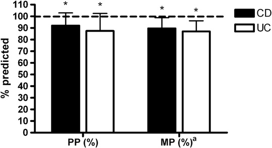
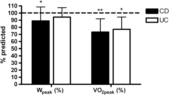
Results Pediatric patients with inflammatory bowel disease had lower PP ($90% of predicted), MP ($88% ofpredicted), Wpeak ($91% of predicted), and VO2peak ($75% of predicted) compared with reference values. When pa-tients with CD or UC were compared separately to reference values, Wpeak was significantly lower only in the CDgroup. No statistically significant correlations were found between any exercise variables and disease duration (r= 0.01 to 0.14, P = .47 to .95) or disease activity (r = -0.19 to -0.31, P = .11 to .38), measured by pediatric CD activityindex or pediatric ulcerative colitis activity index. After controlling for chronological age, recent hemoglobin levelswere significantly correlated with PP (r = 0.45, P = .049), MP (r = 0.63, P = .003), VO2peak (r = 0.62, P = .004), andWpeak (r = 0.70, P = .001).
Conclusions Pediatric patients with inflammatory bowel disease exhibit impaired aerobic and anaerobic exercisecapacity compared with reference values. (J Pediatr 2011;158:814-9).
Inflammatoryboweldisease(IBD)describesinflammatoryconditionsofthesmallandlargeintestines,mostoftendiag- nosed in the second and third decade of life with about a quarter of new cases occurring in children and adoleThe prevalence of IBD among children and youth in Canada has recently been estimated to be between 18 and 70 per 100 000 (depending on geographical location), making this condition a significant public health issue.
The primary disease manifestations for IBD in children and adults do not differ extensively. However, the major difference is that children are at particular risk for extra intestinal manifestation including growth failure, weight loss, and anemia. The diseasecan have a great impact on a child’s self-esteem, as puberty can be delayed.In addition, these effects can be even more profound inchildren with Crohn’s disease (CD) compared with ulcerative colitis (UC), for which anemia and growth failure are not common.
Epidemiologic studies report that adults with IBD tend to lead a sedentary lifestyl– a choice that may lead to reduced exercise capacity, which is an important predictor of mortalityIn adults with CD, for example, aerobic fitness – a key measure of exercisecapacity – was significantly lower than values for healthy adults, suggesting a need for these patients to participate in regular physicalactivity.The extent to which exercise capacity is reduced in pediatric patients with IBD and therefore the extent to which physicalactivity should be counseled with these patients is unknown. It is also important to understand exercise capacity in patients with CDand UC to distinguish possible effects of disease type, given the differences between these conditions in anemia and growth failure.
As part of usual care, patients with IBD from our children’s hospital performed exercise tests at the Children’s Exercise and Nutrition Centre, creating the opportunity to retrospectively analyze fitness data on these patients. The aim of this study was toreport anaerobic and aerobic exercise capacity in pediatric patients with IBD and to determine whether there are differencesbetween patients with CD and UC.
Twenty-nine patients with IBD from the pediatric gastroenterology clinic of the McMaster Children’s Hospital performedexercise tests to assess general fitness levels. All testing was conducted by the same clinical exercise physiologist according to From the Institute of Human Movement Sciences, University of Groningen, Groningen, The Netherlands (H.P.); Wilhelmina Children’s Hospital, University MedicalCenter Utrecht, Utrecht, The Netherlands (T.T.); Children’s Exercise and Nutrition Centre (H.P., T.T., B.W., S.S., B.T.), Department of Pediatrics and PediatricGastroenterology and Nutrition (R.I., R.S.), McMaster The authors declare no conflicts of interest.
0022-3476/$ - see front matter. Copyright standard protocols. The results of 19 patients with CD and 10 lian childrebecause Canadian WC percentiles are not patients with UC are included in this study, which was ap- available for this age group. Z-scores for BMI were calcu- proved by the Hamilton Health Sciences/Faculty of Health lated according to the Centers for Disease Control. Anthro- Sciences Research Ethics Board. Both groups had similar pometric measures and chronological age were used to characteristics (). Disease activity was assessed by estimate years from peak height velocity (PHV) according the pediatric CD (PCDAI) or ulcerative colitis (PUCAI) to Mirwald et this value was used as a marker of bio- disease activity index.Patients were in remission or had mild disease (PCDAI; mean = 6.71, range = 0 to 20 andPUCAI; mean = 0.50, range = 0 to 5). The mean disease duration in patients with CD was 2.5 Æ 1.9 years and in After the anthropometric assessment, anaerobic power of the patients with UC 3.7 Æ 2.55 years. Time spent in remission legs was measured using the Wingate Anaerobic Cycling Test since the last flare-up was available in 14 patients with CD (WAnT) performed on a cycle ergometer (Metabo-Fleisch, and 8 patients with UC and was, respectively, 0.8Æ0.9 and Basel, Switzerland).The WAnT was preceded by a 3-min- 1.3 Æ 1.1 years (not significantly different based on ute warm-up exercise (cycling at a low resistance). The independent t test; P = .23). Information on medication warm-up was interspersed with short sprints at maximal intake was available in 23 patients (6 CD missing), all speed. After 5 minutes of rest or when the heart rate (HR) re- patients were receiving medication (CD/UC: 5-aminosalicylic turned to resting level (whichever came first), the test was acid 10/8, azathioprine 6MP 7/4, methotrexate 2/1, flagyl 4/2, started, with the patient pedaling at maximal speed for 30 infliximab 2/1, prednisone 9/4, entocort 0/1). All prednisone- seconds against a braking force (ie, resistance) set at 45 exposed children were treated starting with 40 mg of g$kgÀ1 body mass. Two performance variables were calcu- prednisone per day; the dose was then decreased by 5 mg per lated: peak mechanical power (PP), which is the highest week over 8 weeks. At the time of exercise testing, recent power output (in Watts) over any 3-second period, and hemoglobin concentrations were available in 22 patients (14 mean mechanical power (MP), which is the average power CD and 9 UC patients) and were 129 Æ 17 g/L and 133 Æ 8 output (in Watts) over the 30-second test. The fatigue index was also calculated as the difference between PP and the low-est power during the test divided by PP and expressed as Height, sitting height (Harpender wall-mounted Stadiome-ter 2109, CMS Weighing Equipment, Ltd, London, United Kingdom), waist circumference (WC, measured 4 cm above Approximately 40 minutes after the WAnT, each patient the naval using a standard anthropometry tape), body mass performed a graded exercise test on the same cycle ergom- (Mott electronic scale, model LC 2424, 20-g accuracy, Santa eter (Metabo-Fleisch) to determine peak oxygen uptake Rosa, California) and percentage body fat by bioelectrical (VO2peak). The McMaster All-Out Progressive Continuous impedance (InBody520, Biospace Co, Ltd, Seoul, Korea) Cycling Test was used with the initial work load and incre- were assessed. Body mass index (BMI) was calculated as ments based on the patient’s height.Work load was in- weight O height2, BMI percentiles were calculated using creased every 2 minutes until the patient could no longer Centers for Disease Control growth charts, and WC percen- pedal at the prescribed cadence (50 rpm), despite strong tiles were calculated using reference values from 11- to 18- encouragement. During the test, expired gas concentrations year-old Canadian childrand 9- to 10-year-old Austra- THE JOURNAL OF PEDIATRICS  www.jpeds.com Anaheim, California). Minute ventilation and O2 uptake were by this patient. One girl with CD could not tolerate the calculated and registered at intervals of 20 seconds. Heart rate mouthpiece, therefore VO2peak could not be determined.
was measured continuously using a Polar Vantage XL HR All other patients completed both exercise tests without issue, monitor (Polar Electro OY, Kempele, Finland). The following and there were no unexpected events during the testing. The performance variables were calculated: VO2peak (L$minÀ1 results of the anaerobic and aerobic exercise test (both pre- and mL$kg body massÀ1$minÀ1) taken as the highest 20-sec- sented as mean Æ standard deviation and as a percent of pre- ond value, peak aerobic mechanical power (Wpeak in Watts dicted for age- and sex-matched values) are shown in and Watts$kg body massÀ1) taken as the last work load achieved and prorated if the full 2-minute stage was not com-pleted, and peak HR (HR highest HR. The clinical protocol to have the aerobic fitness As a group, anaerobic power was significantly lower in the test follow the anaerobic fitness test is the standard protocol patients compared with reference values and were 90 and in our clinic, used for patients with cystic fibrosis and those 89% predicted for PP (Watts$kgÀ1) and MP (Watts$kgÀ1), recovering from cancer, and is also used by other clinics for respectively. No significant differences between patients with CD and UC were found. Given the more than 4 kg ofdifference in fat-free mass (FFM) between patients with CD and UC (albeit nonsignificant), we compared exercise values Peak power, MP, and Wpeak values were compared with refer- normalized for FFM, but no significant differences between ence data from age- and sex-matched healthy youth provided the groups were found for PP (P = .10) or MP (P = .20).
by Bar-Or and RowlanVO2peak values were compared with Given the muscle wasting effects of corticosteroids, we also data from age- and sex-matched healthy youth tested in our compared patients using versus patients not using predni- laboratory. To account for the generally smaller size of patients sone, but we did not find significant differences for PP (P = with IBD, we also made VO2peak comparisons based on age, .72) or MP (P = .19). The fatigue index was not different be- sex, and height; however, the results were identical to match- tween patients with CD and UC (P = .51).
ing for age and sex alone, and the former results (age- and sex-matched) are therefore presented. Using reference data gener- ated with the same protocols, a percentage of predicted value The average HRpeak was 186 Æ 10 beats$minÀ1 for patients was calculated for each exercise variable. Data were normally with CD and 189 Æ 12 beats$minÀ1 for patients with UC. RE- distributed and parametric tests were used when n was $10.
Rpeak averaged 1.15 Æ 0.05 in patients with CD and 1.16 Æ Differences between patients with CD and UC were tested us- 0.09 in patients with UC. These values for HRpeak and RE- ing independent-samples t tests. Differences between patients Rpeak confirm that patients provided a maximal effort.
and reference data were compared using paired-samples t tests As a group, indices of aerobic power were significantly or Wilcoxon signed-rank tests when n was <10. Associations lower in patients compared with reference values and were between variables were tested using Pearson correlation coef- 91%, 75%, and 79% predicted for Wpeak (Watts$kgÀ1) and ficients. An a level of <0.05 was considered as statistically sig- (L$minÀ1 and mL$kgÀ1$minÀ1), respectively. When patients nificant. All analyses were performed using SPSS (SPSS Inc, with CD or UC were compared separately with reference values, Wpeak was significantly lower only in the patientswith CD. However, when actual values from the anaerobic and aerobic tests were compared between patients with CDand UC, no significant group differences emerged. When One boy with CD felt unwell after the WAnT (nausea and values were normalized for FFM, no significant group differ- vomiting); therefore no aerobic power test was performed ences were found for Wpeak (P = .20) or VO2peak (P = .19).
Table II. Anaerobic and aerobic exercise measurements *P < .05 compared with reference values.
†P < .001 compared with reference values.
zLevel of significance based on CD, n = 11; UC, n = 7.
ter controlling for age, hemoglobin remained significantlycorrelated with PP (Watts$kg-1; r = 0.45, P = .049), MP(Watts$kgÀ1; r = 0.63, P = .003), Wpeak (Watts$kgÀ1; r =.70, P = .001), VO2peak (L$min–1; r = 0.60, P = .006), andVO2peak (mL$kgÀ1$min–1; r = 0.62, P = .004).
To determine whether patients who performed well on the WAnT also performed well on the test of aerobic power, cor-relational analysis was performed on these variables andfound positive and significant relationships for all exercisevariables (r = 0.45 to 0.85, P = .023 to .001). Given the impor-tance of muscle mass for exercise performance, we also deter- Figure 1. Anaerobic exercise variables as percent predicted mined the association between FFM and exercise variables.
(Æ SD). Dashed line is 100%; *P < .05 compared with 100%; There was a strong association between FFM and PP areference data based on children younger than 15 years; (Watts$kgÀ1; r = 0.66, P = .001), MP (Watts$kgÀ1; r = Crohn disease, n = 11; ulcerative colitis, n = 7.
0.68, P = .001) and Wpeak. (Watts$kgÀ1; r = 0.48, P = .010),and (L$min–1; r = 0.93, P = .001).
Comparisons between patients using prednisone and not us- ing prednisone revealed a significant difference for Wpeak(P = .048) but not for VO Although impaired exercise capacity has been reported in adults with IBD, the extent of this problem in pediatric pa- tients is unknown. We report that measures of both anaero- To determine whether disease duration and disease activity bic and aerobic capacity (PP, MP, Wpeak, and VO2peak,) were (measured by PCDAI or PUCAI) were associated with our significantly lower in pediatric patients with IBD compared indices of exercise capacity, Pearson correlational analysis with reference values from healthy youth, which is in accor- was performed. No statistically significant correlations were dance with previous results for aerobic capacand anaer- found between any exercise variable and disease duration (r obic capacityin adults with CD. Patients with IBD that = 0.01 to 0.14, P = .47 to .95) or disease activity (r = À0.19 showed lower aerobic capacity were most likely also to to À0.31, P = .11 to .38). Hemoglobin concentrations, which show low anaerobic capacity, suggesting a general deficiency might serve as a disease activity variable in patients with in exercise capacity and not an impairment in one body sys- IBD,was found to be below the age- and sex-based labora- tem (eg, cardiovascular) over another (eg, skeletal muscle).
tory reference values in 5 of 22 patients (4 CD and 1 UC). He- Compared with reference values, aerobic fitness was moglobin correlated with PP (Watts$kgÀ1; r = 0.50, P = slightly lower in patients with CD than UC; however, differ- .014), MP (Watts$kgÀ1; r = 0.65, P = .001), VO ences in actual values were not statistically significant be- tween the two disease groups. Additional post hoc calculations based on our data revealed that at least 100 pa- .001). No statistically significant difference in hemoglobin tients in each group would have to be tested for these results was found between patients with CD and UC. To control to be statistically significant with a statistical power of 80%.
for the possible influence of chronological age on hemoglo- In light of these estimations, it is likely that the clinical ap- bin, Pearson partial correlational analysis was performed. Af- proach to pediatric patients with CD or UC would nothave to be different in terms of exercise testing. That aerobicfitness, as measured by the gold standard VO2peak, was lowerin youth with CD ($75% of predicted) is worrisome becauseexercise capacity is a strong indicator for the development offuture cardiovascular disease risk factorThere is strongscientific evidence that youth with low aerobic fitness aremore likely to display additional risk factors for cardiovascu-lar disease such as elevated blood pressure.Our new find-ings in youth with IBD suggest that their reduced aerobicfitness make them at even greater risk for cardiovascular dis-ease than their peers without IBD and that interventions areneeded.
One possible explanation for this lower-than-predicted ex- ercise capacity might be the low hemoglobin concentrations Figure 2. Aerobic exercise variables as percent predicted in the patients. We found a significant correlation between (Æ SD). Dashed line is 100%; * P < .05 compared with 100%; the aerobic fitness indices and hemoglobin in those patients **P < .001 compared with 100%.
for whom recent hemoglobin levels were available. Low Exercise Capacity in Pediatric Patients with Inflammatory Bowel Disease THE JOURNAL OF PEDIATRICS  www.jpeds.com hemoglobin concentrations are a common problem in IBD.
for clinical outcomes and quality of life, as reported for other It is related to a loss of weight, low tolerance to the underlying disease, and a poor growth rate in childrenThus, the issue A limitation of this study is that we did not test the com- of possible anemia should be more closely investigated and plete patient population at our children’s hospital. Moreover, iron supplementation, when warranted, may be one strategy reference values were unavailable for MP values in children to help improve exercise endurance in these patients.
older than 15 years. Therefore, our results may not be appli- It is also possible that a lower exercise capacity could be re- cable to the general pediatric IBD population, particularly lated to increased baseline levels of inflammatory mediators those with active disease. A prospective study investigating (eg, tumor necrosis factor-a) that characterize IBD. Tumor exercise capacity and its association with hemoglobin levels necrosis factor-a is associated with cachexia, which can and inflammation in pediatric patients with IBD is war- lead to lower levels of aerobic capacity independent of the ranted. In addition, the treatment of low hemoglobin con- type of inflammatory diHigh baseline levels of pro- centrations with iron supplementation, when warranted, inflammatory cytokines in patients with chronic heart failure might potentially improve exercise capacHowever, and chronic obstructive pulmonary disease have been associ- this has not yet been studied in pediatric patients with IBD.
ated with an impaired exercise capacity and low anaerobic Pediatric patients with IBD exhibited impaired aerobic and aerobic capacity independent from the primary disease and anaerobic exercise capacity even when in remission location (eg, impaired heart or lung funcAlthough from their disease. Clinicians who specialize in pediatric we did not measure inflammation in our patients, resting IBD should consider referring patients with IBD for exercise levels of pro-inflammatory cytokines in patients both in re- testing and involvement of an activity therapist or physio- mission and with active disease have been found to be ele- therapist in the clinical management of these children and vatedand might therefore be partly an explanation for the lower then predicted anaerobic and aerobic exercisevalues.
Submitted for publication Dec 16, 2009; last revision received Aug 31, 2010; In this study, we found evidence that medication use could contribute to low levels of exercise capacity. Because Reprint requests: Dr Brian W. Timmons, PhD, Children’s Exercise and NutritionCentre, McMaster University and McMaster Children’s Hospital, Chedoke corticosteroids are known for their myopathic effewe Hospital, Evel Bldg, Room 469, Sanatorium Road, PO Box 2000, Hamilton, ON, performed a comparison between patients using corticoste- roids and patients not using corticosteroids. Aerobic capacitytended to be lower in patients using corticosteroids (predni- sone), with a significant difference in Wpeak. Recent stud-in youth with CD showed persistent deficits in lean 1. Saibil FG. Crohn’s Disease and Ulcerative Colitis. Toronto: Key Porter tissue, with corticosteroid use and chronic inflammation as possible mechanisms responsible for these deficits. In our pa- 2. Kim SC, Ferry GD. Inflammatory bowel diseases in pediatric and adoles- tients, FFM did not differ significantly from reference values cent patients: clinical, therapeutic, and psychosocial considerations.
Gastroenterology 2004;126:1550-60.
nor were differences seen between patients on and off corti- 3. Griffiths AM. Specificities of inflammatory bowel disease in childhood.
costeroids (results not shown), suggesting that impaired ex- Best Pract Res Clin Gastroenterol 2004;18:509-23.
ercise capacity in these patients may not be related to relative 4. Bernstein CN, Blanchard JF, Rawsthorne P, Wajda A. Epidemiol- ogy of Crohn’s disease and ulcerative colitis in a central Canadian Exercise training and the promotion of daily physical ac- province: a population-based study. Am J Epidemiol 1999;15(149):916-24.
tivity seem indicated in pediatric patients with IBD. How- 5. Kelsen J, Baldassano RN. Inflammatory bowel disease: the difference be- ever, no information exists about the effects of acute or tween children and adults. Inflamm Bowel Dis 2008;14(Suppl 2):S9-S11.
chronic exercise on the underlying inflammation associated 6. Narula N, Fedorak RN. Exercise and inflammatory bowel disease. Can J with the pathology of High-intensity exercise might lead to gastrointestinal ischemiabecause splanchnic blood 7. Blair SN, Kohl HW, III, Paffenbarger RS, Jr., Clark DG, Cooper KH, Gibbons LW. Physical fitness and all-cause mortality: a prospective study flow is reduced during exercise compared with resting condi- of healthy men and women. JAMA 1989;262:2395-401.
tions.Besides enhancing exercise capacity, exercise training 8. Loudon CP, Corroll V, Butcher J, Rawsthorne P, Bernstein CN. The ef- might be a possible anabolic stimulus to decrease levels of in- fects of physical exercise on patients with Crohn’s disease. Am J Gastro- flammation and might therefore also have a positive effect on growth factors (eg, IGF-I).To date, several randomized, 9. Hyams JS, Ferry GD, Mandel FS, Gryboski JD, Kibort PM, Kirschner BS, et al. Development and validation of a pediatric Crohn’s disease activity controlled studies in various pediatric patient groups have index. J Pediatr Gastroenterol Nutr 1991;12:439-47.
shown improvements in exercise capacity in children with 10. Turner D, Otley AR, Mack D, Hyams J, de BJ, Uusoue K, et al. De- conditions ranging from cerebral palsy to cystic fibrosis.
velopment, validation, and evaluation of a pediatric ulcerative colitis The optimal training paradigm (sufficient efficacy without activity index: a prospective multicenter study. Gastroenterology adverse effects on disease activity) for youth with IBD re- 11. Katzmarzyk PT. Waist circumference percentiles for Canadian youth 11- mains to be determined. However, our findings indicate 18 y of age. Eur J Clin Nutr 2004;58:1011-5.
that close clinical monitoring of markers of fitness seems 12. Eisenmann JC. Waist circumference percentiles for 7- to 15-year-old warranted in youth with IBD and may serve as a surrogate Australian children. Acta Paediatr 2005;94:1182-5.
13. Mirwald RL, Baxter-Jones AD, Bailey DA, Beunen GP. An assessment of 25. Kmiec Z. Cytokines in inflammatory bowel disease. Arch Immunol Ther maturity from anthropometric measurements. Med Sci Sports Exerc 26. Sanchez-Munoz F, Dominguez-Lopez A, Yamamoto-Furusho JK. Role 14. Bar-Or O, Rowland TW. Pediatric Exercise Medicine: From Physiologic of cytokines in inflammatory bowel disease. World J Gastroenterol Principles to Health Care Applications. Champaign, IL: Human Kinet- 27. Kader HA, Tchernev VT, Satyaraj E, Lejnine S, Kotler G, Kingsmore SF, 15. van Brussel M, van Doren L, Timmons BW, Obeid J, van der Net J, et al. Protein microarray analysis of disease activity in pediatric inflamma- Helders PJ, et al. Anaerobic-to-aerobic power ratio in children with ju- tory bowel disease demonstrates elevated serum PLGF, IL-7, TGF-beta1, venile idiopathic arthritis. Arthritis Rheum 2009;15(61):787-93.
and IL-12p40 levels in Crohn’s disease and ulcerative colitis patients in re- 16. Mack DR, Langton C, Markowitz J, LeLeiko N, Griffiths A, Bousvaros A, mission versus active disease. Am J Gastroenterol 2005;100:414-23.
et al. Laboratory values for children with newly diagnosed inflammatory 28. Schakman O, Gilson H, Kalista S, Thissen JP. Mechanisms of muscle at- bowel disease. Pediatrics 2007;119:1113-9.
rophy induced by glucocorticoids. Horm Res 2009;72(Suppl 1):36-41.
17. Brevinge H, Berglund B, Bosaeus I, Tolli J, Nordgren S, Lundholm K. Ex- 29. Sylvester FA, Leopold S, Lincoln M, Hyams JS, Griffiths AM, Lerer T. A ercise capacity in patients undergoing proctocolectomy and small bowel two-year longitudinal study of persistent lean tissue deficits in children resection for Crohn’s disease. Br J Surg 1995;82:1040-5.
with Crohn’s disease. Clin Gastroenterol Hepatol 2009;7:452-5.
18. Wiroth JB, Filippi J, Schneider SM, Al-Jaouni R, Horvais N, Gavarry O, 30. Thayu M, Denson LA, Shults J, Zemel BS, Burnham JM, Baldassano RN, et al. Muscle performance in patients with Crohn’s disease in clinical re- et al. Determinants of changes in linear growth and body composition in mission. Inflamm Bowel Dis 2005;11:296-303.
incident pediatric Crohn’s disease. Gastroenterology 2010;139:430-8.
19. Carnethon MR, Gidding SS, Nehgme R, Sidney S, Jacobs DR, Jr., 31. Ploeger HE, Takken T, de Greef MH, Timmons BW. The effects of acute Liu K. Cardiorespiratory fitness in young adulthood and the de- and chronic exercise on inflammatory markers in children and adults velopment of cardiovascular disease risk factors. JAMA 2003;290: with a chronic inflammatory disease: a systematic review. Exerc Immu- 20. Tolfrey K, Campbell IG, Jones AM. Selected predictor variables and the 32. Kolkman JJ, Groeneveld AB, van der Berg FG, Rauwerda JA, lipid-lipoprotein profile of prepubertal girls and boys. Med Sci Sports Meuwissen SG. Increased gastric PCO2 during exercise is indicative of gastric ischaemia: a tonometric study. Gut 1999;44:163-7.
21. Christodoulou DK, Tsianos EV. Anemia in inflammatory bowel disease: 33. Rowell LB. Human Cardiovascular Control. Oxford: Oxford University the role of recombinant human erythropoietin. Eur J Intern Med 2000; 34. Street ME, de’Angelis G, Camacho-Hubner C, Giovannelli G, Ziveri MA, 22. Roubenoff R. Exercise and inflammatory disease. Arthritis Rheum 2003; Bacchini PL, et al. Relationships between serum IGF-1, IGFBP-2, interleukin-1beta and interleukin-6 in inflammatory bowel disease.
23. Skeletal muscle dysfunction in chronic obstructive pulmonary disease. A statement of the American Thoracic Society and European Respiratory 35. Klijn PH, Oudshoorn A, Van der Ent CK, van der NJ, Kimpen JL, Society. Am J Respir Crit Care Med 1999;159(4 Pt 2):S1-40.
Helders PJ. Effects of anaerobic training in children with cystic fibrosis: 24. Nessler J, Nessler B, Kitlinski M, Gackowski A, Piwowarska W, a randomized controlled study. Chest 2004;125:1299-305.
Stepniewski M. Concentration of BNP, endothelin 1, pro-inflammatory 36. Mann SK, Kaur S, Bains K. Iron and energy supplementation improves cytokines (TNF-alpha, IL-6) and exercise capacity in patients with heart the physical work capacity of female college students. Food Nutr Bull failure treated with carvedilol. Kardiol Pol 2008;66:144-51.
Exercise Capacity in Pediatric Patients with Inflammatory Bowel Disease

Source: http://www.hildeploeger.nl/Ploeger_(2011)_Exercise_capacity_in_pediatric_patients_with_IBD.pdf

math.utoledo.edu

Department of MathematicsThe University of Toledo2801 W. Bancroft St Toledo, Ohio 43606-3390Tel:(O)(419) 530-2998 Fax: (419) 530-4720Email:[email protected]:http://www.math.utoledo.edu/∼mtsuiDifferential Geometry (53), General Relativity and Gravitation (83) andPartial Differential Equations (35). • Ph. D. in Mathematics, 2001, Brandeis University• M.S. in Applied Mathemat
drevensek.si

CALINIC Y The science of photodisinfection has a fascinating treatment alternative as a potential tool, and then history. Literature dates back to 1500 BC decide which of these tools are best suited for a describing therapy involving the placement of certain specific problem. A patient may benefit most from kinds of seeds on the skin which were then exposed to a “conservative” or

Copyright © 2013-2018 Pharmacy Abstracts